ARTICLE

김치에서 분리한 Leuconostoc mesenteroides가 생산하는 Bifidobacterium 성장 자극 물질의 분리 및 특성

권윤경1https://orcid.org/0009-0005-4019-7279, 박재용1,*https://orcid.org/0000-0002-1088-3930
Yunkyoung Kwon1https://orcid.org/0009-0005-4019-7279, Jae-Yong Park1,*https://orcid.org/0000-0002-1088-3930
Author Information & Copyright
1대구가톨릭대학교 식품영양학과
1Department of Food Science and Nutrition, Daegu Catholic University, Gyengsan 38430, Republic of Korea
*Corresponding author : Jae-Yong Park, Department of Food Science and Nutrition, Daegu Catholic University, Gyengsan 38430, Republic of Korea, Tel : 82-53-850-3521, E-mail : jaepark@cu.ac.kr

© Copyright 2023 Resource Science Research Institute, Kongju National University. This is an Open-Access article distributed under the terms of the Creative Commons Attribution Non-Commercial License (http://creativecommons.org/licenses/by-nc/4.0/) which permits unrestricted non-commercial use, distribution, and reproduction in any medium, provided the original work is properly cited.

Received: Nov 20, 2023; Revised: Dec 15, 2023; Accepted: Dec 18, 2023

Published Online: Dec 31, 2023

Abstract

Genus Bifidobacterium is known to be beneficial gut bacteria, and some strains of Bifidobacterium are being used as probiotics. To find lactic acid bacteria (LAB) that produce Bifidobacterium growth stimulator (BGS), which promotes the growth of Bifidobacterium, we used 733 strains isolated from kimchi for BGS screening and selected strain K748, which showed the highest BGS activity at 320 AU/mL. The strain was identified as Leuconostoc mesenteroides by 16s rDNA sequencing, and the strain was named L. mesenteroides K748. BGS activity increased from 12 hours after incubation in MRS medium, reaching maximum activity (320 AU/mL) at 15 hours and maintaining that activity. The ethyl acetate extract of L. mesenteroides K748 culture supernatant promoted the growth of Bifidobacterium longum, and HPLC using this extract revealed a peak with the same retention time (7.5 minutes) as 1,4-dihydroxy-2-naphthoic acid (DHNA). LC-MS analysis revealed that the DHNA and ethyl acetate extracts of L. mesenteroides K748 had the same molecular weight, 204.1. Overall, it is possible to conclude that L. mesenteroides K748 produces DHNA that promotes the growth of Bifidobacterium.

Keywords: Bifidogenic growth stimulator; Leuconostoc mesenteroides; Kimchi

I. 서 론

인간의 장에는 약 100조 이상의 장내 미생물 세포가 서식하고 있으며, 이들은 Firmicutes 문, Bacteroidetes 문, Actinobacteria 문, α-Proteobacteria 문 등에 해당한다(Doré and Blottière, 2015; Khanna and Tosh, 2014). 장내 미생물 균총은 유익균이 유해균보다 많은 상태일 때 균형을 이루고, 반대로 유해균이 증가하면 불균형 상태로 변화되며, 이러한 미생물 균총의 변화는 식이, 노화, 유전, 면역, 스트레스 등 내·외부 요인의 영향을 받는다(Meyer and Stasse-Wolthuis, 2009). 현대인들의 고지방·고당류 식사와 같은 서구화된 식습관 및 비활동적 생활방식, 항생제 남용 등은 장내 미생물의 불균형을 초래하며, 이로 인해 비만, 당뇨, 고혈압 등과 같은 대사성 질환과 염증성 장 질환, 과민성 대장 증후군과 같은 위장관 질환, 심혈관계 질환 등이 초래된다. 반면, 균형 잡힌 식습관 및 생활방식, 프로바이오틱스 섭취 등으로 인해 장내 유익균이 증가하면 장내 미생물은 균형을 이루고, 인간에게 생리학적 이점을 제공할 수 있다(Kim, 2013; Knysh, 2019; Nagpal et al., 2014).

최근, 장내 미생물에 대한 관심이 높아짐에 따라 프로바이오틱스 및 프리바이오틱스의 사용이 증가하고 있다. 프로바이오틱스는 적절한 양을 섭취할 때, 인간에게 건강상 이점을 주는 살아있는 미생물로 정의되며, 일반적으로 Lactobacillus 속, Bifidobacterium 속이 이에 속한다(Iraporda et al., 2019). 프리바이오틱스는 프로바이오틱스의 성장 및 활동을 유도하는 비소화성 식품 성분으로, 대표적으로 이눌린, 프락토올리고당(FOS), 갈락토올리고당(GOS) 등을 포함한다. 신바이오틱스는 프로바이오틱스와 프리바이오틱스를 결합한 식품 성분 및 보충제이며, Bifidobacterium과 프락토올리고당, Lactobacillus rhamnosus GG와 이눌린을 결합한 것을 예로 들 수 있다(Gourbeyre et al., 2011; Pandey et al., 2015). 포스트바이오틱스는 프로바이오틱스에 의해 생산 및 분비되는 대사산물 및 부산물로 효소, 단백질 및 펩타이드, 유기산, 단쇄지방산(SCFA), 비타민 B, 비타민 K 등이 있다(Aguilar-Toalá et al., 2018). 프로바이오틱스와 포스트바이오틱스는 장내 pH 감소, 항균물질 생산, 상피 장벽 강화, 면역계 조절, 경쟁적 배제작용 등을 통해 병원성 미생물의 성장 및 유착을 저해시키고, 미생물 균총을 균형적으로 변화시키며, 항염증, 면역 향상, 장 질환 개선, 배변 활동 개선 등 생리학적으로 유익한 효과를 나타낸다(Iraporda et al., 2019; Meyer and Stasse-Wolthuis, 2009; Piqué et al., 2019).

Bifidobacterium은 장내 미생물 균총 중 가장 유익한 균으로 인식되고 있으며 병원성 균에 대한 길항작용, 비타민 B군 생산, 유당불내증 개선 효과, 면역 증강, 항종양 등 역할을 하는 것으로 알려져 있다(Jung et al., 2020; Park and Ji, 2010; Vitellio et al., 2019). B. bifidum BGN4 및 B. longum BORI의 경우 뮤신 분해능이 없고, 암모니아나 생체 아민 등 유해 물질을 거의 생산하지 않으며, 독성 유전자를 가지지 않는다는 보고가 있으며(Kim et al., 2018), B. longum BB536의 경우 체내 항상성 조절 및 항알레르기 효과를 가지는 것으로 보고되었다(Wong et al., 2019).

Bifidobacterium 속 균주의 성장을 자극시키는 프리바이오틱스를 Bifidogenic growth stimulator (BGS)라고 부르며, 모유 및 우유 성분에 속하는 κ-카제인, 카제이노마이크로펩타이드, 다당류에 속하는 이눌린, 올리고당류에 속하는 FOS, GOS, 라피노오스, 스타키오스, 락툴로오스, 당알코올류에 속하는 락티톨, 퀴논류에 속하는 2-amino-3-carboxy-1,4-naphthoquinone (ACNQ)과 1,4-dihydroxy- 2-naphthoic acid (DHNA) 등으로 분류할 수 있다(Nagpal et al., 2014). 이눌린 및 올리고당을 첨가한 탈지유는 Bifidobacterium 속 균주의 성장 및 활성, 생존율 증가에 영향을 미치고(Choi and Shin, 2006), FOS 및 GOS가 포함된 배지에서 Bifidobacterium 속 균주를 배양했을 때, Bifidobacterium의 성장이 유의적으로 증가되었으며(Sims and Tannock, 2020), 락툴로오스를 경구 섭취한 인간의 대변에서 Bifidobacteium 속의 균수와 비율이 증가하였다는 연구가 보고된 바 있다(Sakai et al., 2019). 이러한 연구를 바탕으로 이눌린 및 올리고당과 같은 프리바이오틱스는 현재 상업적으로 판매가 되고 있다. 퀴논류 BGS인 ACNQ 및 DHNA는 발효식품에서 유래된 유산균에서 생산되었다는 연구 결과가 다수 보고되었다. 그 예로, ACNQ는 치즈에서 분리한 Propionibacterium freudenreichii 7025(Mori et al., 1997), DHNA는 치즈에서 분리한 P. freudenreichii ET-3(Isawa et al., 2002), Lactobacillus casei LP1(Kang et al., 2015), Lactobacillus plantarum LP2(Kim and Moon, 2015) 등에서 생산된다고 알려져 있으며, 상업적 막걸리에서 DHNA가 검출되기도 하였다(Eom et al., 2012).

현재, Bifidobacterium 성장 자극을 나타내는 BGS와 관련된 많은 연구가 보고되고 있으나, 김치 유산균을 이용한 연구는 부족한 실정이다. 본 연구는 유산균을 활용하여 Bifidobacterium 속 균주의 성장을 높이는 방법을 찾고자 하였으며, Bifidobacterium 성장에 필요한 BGS를 생산하는 유산균을 한국의 전통 발효식품인 김치에서 분리하고, 그 특성을 규명하고자 한다.

Ⅱ. 재료 및 방법

유산균 분리

식당 및 일반 가정용 김치 21종을 유산균 분리에 사용하였다. 김칫국물을 단계 희석하여 BCP 한천 배지(20 g tryptone, 5 g yeast extract, 4 g NaCl, 1.5 g sodium acetate, 0.04 g bromocresol purple, 15 g agar per liter)에 도말한 후 30°C에서 24시간 배양하였다. 노란색 환을 생성하는 집락을 MRS 액체 배지(BD DifcoTM, USA)에 접종하여 30°C에서 24시간 배양한 후, 한 번 더 계대 배양하였고, 80% glycerol stock을 제조하여 –70°C에 보관하였다.

BSG 생산 유산균 탐색

MRS 액체 배지에 30°C, 24시간 배양한 균주 배양액을 원심분리기(1730MR, Gyrozen, Korea)를 이용해 원심분리(14,000 × g, 3 min)하여 상등액만 회수하였다. 회수한 상등액을 0.2 μm Syringe filter (Cellulose acetate, Hydrophilic, Sartorius, Germany)를 이용해 여과하였고, –70°C에서 보관하였다가 실험에 사용하였다. BGS 활성이 높은 균주를 선발하기 위해 앞서 준비한 균주 상등액을 이용하여 스크리닝을 실시하였다. BGS 활성 측정에 지표 균주로 사용된 Bifidobacterium longum subsp. longum KCTC 3128 (이하, B. longum KCTC 3128)는 생물자원센터(KCTC)에서 분양받았고, RCM 액체 배지(BD DifcoTM, USA)에 접종하여 37°C에서 24시간 동안 혐기적(BD Gaspak EZ™ Container Systems, BD DifcoTM, USA)으로 배양하여 실험에 사용하였다.

스크리닝은 Kouya 등의 방법을 변형한 한천배지확산법으로 2번에 걸쳐 진행하였다 (Kouya et al., 2007). 멸균 후 식힌 TPY 한천 배지(30 g glucose, 8 g trypticase peptone, 3 g phytone peptone, 5 g yeast extract, 2 g K2HPO4, 3 g KH2PO4, 0.5 g MgCl2·6H2O, 0.5 g L-cysteine, 0.01 g FeSO4·7H2O, 15 g agar per liter, pH 8.5)에 10배 희석한 B. longum KCTC 3128 배양액을 1% 접종한 다음, 페트리접시에 20 mL씩 분주하고 굳힌 후에 멸균된 8 mm 디스크(Advantec Toyo Kaisha, Japan)를 배지 표면에 배치하였다. 1차 스크리닝에서는 균주 상등액을 8배 희석하여 사용하였고, 2차 스크리닝에서는 이진희석(2-1-2-8)하여 사용하였다. 희석한 상등액을 배지 표면에 배치된 8 mm 디스크에 50 μL 분주한 후, 37°C에서 24시간 동안 혐기적 배양하여 디스크 주위의 B. longum KCTC 3128 성장환을 통해 BGS 활성 유무를 확인하였다. BSG 활성의 크기는 임의단위(AU)를 사용하였으며, 희석한 배양상등액 50 μL를 가하였을 때 성장환을 나타내는 최고 희석배수의 역수로 정의하였다.

선발 유산균의 동정

최종 선발된 BGS 생산 균주를 MRS 액체 배지에 30°C에서 24시간 배양한 후, 배양액의 chromosomal DNA를 분리하였다. Chromosomal DNA의 16s rDNA gene은 27f(5’-AGAGTTTGATC MTGGCTCAG-3’) 및 1525r(5’-AAGGAGGTGWTCCARCC-3’) primer를 사용하여 PCR (T100TM Thermal Cycler, Bio-rad, Singapore)로 증폭하였다. PCR은 pre-denaturation (94°C, 5 min) 후에 denaturation (94°C, 30 sec), annealing (55°C, 30 sec), extension (72°C, 1 min)을 30회 반복하고, 최종 elongation (72°C, 5 min) 순으로 진행하였다. 증폭된 16s rDNA gene을 pGEM-T easy vector(Promega, USA)에 ligation하고, KCM법을 이용해 E. coli DH5α transformation을 한 후, Exprep TM plasmid kit (GeneAll, Korea)를 이용해 plasmid DNA를 추출하여 DNA 염기서열 분석을 진행하였다. DNA 염기서열 분석은 코스모진텍(COSMO Genetech, Korea)에 의뢰하였으며, ezbiocloud (https://www.ezbiocloud.net/)을 활용하여 동정하였다. API 50 CHL kit(BioMérieux, France)를 이용해 49종의 탄수화물에 대한 발효능을 확인하였다. 실험 방법은 매뉴얼을 참고하였고, 간략히 설명하면 다음과 같다. 최종 선발 균주를 MRS 한천 배지에 30°C, 24시간 배양한 후, 배지 표면의 집락을 회수하여 Suspension medium 2 mL에 현탁하였다. 현탁한 균액을 Suspension Medium 5 mL에 n방울 첨가하여 2 McFarland 용액과 탁도를 비교한 후, API 50 CHL medium에 2n방울 접종하여 실험에 사용하였다. 준비한 시료를 스트립의 튜브에 분주하여 37°C, 24시간 및 48시간 배양 후 결과를 확인하였다.

BSG 활성측정

최종 선발 균주를 MRS 액체 배지에 접종한 후, 30°C에서 배양하면서 UV spectrophotometer (UVmini-1240, Shimadzu, Japan)를 이용하여 24시간 동안 3시간 간격으로 600 nm 파장에서 흡광도를 측정하였으며, 원심분리기(1730MR, Gyrozen, Korea)를 이용해 원심분리(14,000 × g, 3 min)하여 균주 상등액을 회수하였다. 회수한 상등액을 0.2 μm Syringe filter를 이용해 여과하였고, -70°C에서 보관하며 사용하였다. 시간별 상등액의 BGS 활성은 2차 스크리닝에 사용된 한천배지확산법을 이용하여 측정하였다.

BSG 첨가 효과 확인

최종 선발 균주의 2% 배양 상등액을 첨가한 RCM 액체 배지에 미리 배양된 B. longum KCTC 3128을 1% 접종하여 37°C에서 혐기적 배양하면서 15, 18, 21, 24시간마다 UV spectrophotometer를 이용하여 600 nm의 파장에서 배양액의 흡광도를 측정하였다. 상등액을 첨가하지 않은 B. longum KCTC 3128 배양액을 대조군으로 사용하였다. 실험 결과는 3번 반복 측정하였으며, IBM SPSS statistics software 19.0(Statistical Package for Social Sciences, SPSS Inc., Chicago, IL, USA)을 사용하여 분석하였다. 통계적 유의수준은 독립표본 T-검정을 통해 p<0.05로 실시하였다.

High performance liquid chromatography(HPLC)

최종 선발 균주를 MRS 액체 배지에 30°C, 24시간 배양하여 원심분리기(5810R, Eppendorf, Germany)를 이용하여 원심분리(1,500 × g, 20 min)한 후, 0.2 μm Syringe filter를 이용해 여과하였다. 상등액을 hexane, ethyl acetate, butanol 순으로 2번에 걸쳐 분획(supernatant: solvent = 1: 2)하였고, 분획물을 50°C 이하에서 진공감압농축기(EYELA, Tokyo Rikakikai Co., Japan)를 이용하여 농축하였다. 농축된 시료를 ddH2O에 용해하여 흡광도를 이용한 BGS 활성 실험에 사용하였다. 가장 높은 활성을 보인 용매 추출물을 이용해 HPLC 분석을 하였으며, 분석 조건은 Table 1과 같다. 농축된 시료는 methanol(HPLC grade, Merck, Germany)에 용해한 후 사용하였다. 표준물질은 1,4-dihydroxy-2- naphthoic acid(DHNA, Sigma- Aldrich, USA)를 사용하였으며, 농도별로 0, 0.5, 1, 2 μg/mL로 제조하였다. 표준물질을 이용하여 작성한 검량곡선을 통해 시료의 DHNA 함량을 계산하였다.

Table 1. HPLC analysis condition for DHNA
Item Operation condition
HPLC system YL9100 Plus HPLC System (Young In, Korea)
Column TSKgel ODS-100V column phase C18 (4.6 × 150 mm, 5 μm; TOSOH, Japan)
Solvent Solvent A: 0.1% formic acid in water
Solvent B : 0.1% formic acid in acetonitrile
Gradient 0 min (A 80%, B 20%) → 10 min (B 100%) → 13 min (B 100%) → 15 min (A 80%, B 20%) → 20 min (A 80%, B 20%)
Column temperature 40°C
Flow rate 1 mL/min
Injection volume 20 μL
UV detection UV 254 nm
Download Excel Table
Liquid chromatography-mass spectrometry(LC-MS)

최종 선발 균주의 BGS 물질을 확인하기 위해 LC-MS 분석을 진행하였으며 (Eom et al., 2012), 서울대학교 그린바이오공동기기센터에 분석을 의뢰하였다. LC-MS 조건은 Table 2와 같다. 분석 물질의 이온화는 ESI(electrospray ionization) 방법을 사용하여 양이온화 모드로 수행되었으며, MS 스펙트럼은 MRM(multiple reaction monitoring) 모드로 확인하였다. 이온화에 사용된 ESI 조건은 다음과 같다: Nebulizer, 45 psi; gas temperature, 300°C; gas flow, 5 L/min; sheath gas temperature, 250°C; sheath gas flow, 11 L/min; capillary voltage, 3,500 V; nozzle voltage, 500 V.

Table 2. LC-MS analysis condition for DHNA
Item Operation condition
LC-MS system 1290 Infinity II LC system (Agilent, USA)
6470 Triple Quadrupole (QQQ) LC/MS system1) (Agilent, USA)
Column Poroshell 120, EC-C18 (2.1 × 100mm, 2.7 μm; Agilent, USA)
Solvent Solvent A: 0.1% formic acid in water
Solvent B: 0.1% formic acid in acetonitrile
Gradient 0 min (A 90%, B 10%) → 0.5 min (A 90%, B 10%) → 7 min (B 100%) → 13 min (B 100%)
Column temperature 30°C
Flow rate 0.4 mL/min
Injection volume 5 μL
UV detection UV 260 nm

1) QQQ LC-MS system was used for ESI (Electrospray ionization) of isolated compounds.

Download Excel Table

Ⅲ. 결과 및 고찰

BGS 생산 균주 선발

21종의 김치에서 분리한 703개의 유산균 상등액을 이용하여 BGS 활성을 확인하였다(Kouya et al., 2007). 디스크 주변의 B. longum KCTC 3128 성장환이 관찰되면 BGS 활성이 있는 것으로 보고 활성을 계산하여 160 AU/mL 이상의 활성을 보이는 총 22개의 균주를 선발하였다. 1차 선발된 22개 균주 중 가장 높은 BGS 활성을 보이는 균주를 선택하기 위해 2차 스크리닝을 진행하였고, 그 결과는 Table 3과 같다. K748 균주는 320 AU/mL로 2차 스크리닝 균주 중 가장 높은 BGS 활성을 나타냈다. K748 균주를 제외한 21개 균주는 80-160 AU/mL의 BGS 활성을 나타냈다. 따라서, K748 균주를 최종 균주로 선발하였다.

Table 3. 2nd screening of BGS-producing LAB
Strain No. BGS activity (AU/mL)1)
24h2) 48h
K321 80 80
K323 80 160
K345 160 160
K438 80 160
K440 80 160
K443 80 160
K473 160 160
K523 160 160
K560 160 160
K579 160 160
K604 160 160
K633 80 80
K725 160 160
K731 160 160
K736 160 160
K737 80 160
K738 160 160
K743 160 160
K744 160 160
K748 320 320
K783 160 160
K973 160 160

1) The activity per 1 mL sample is defined as 20 AU.

2) Incubation time (hour).

Download Excel Table
선발균주의 동정

최종 균주로 선발된 K748 균주의 16s rDNA 염기서열 분석 결과는 Table 4와 같다. K748 균주는 Leuconostoc mesenteroides와 99% 이상의 유사성을 보였으며, L. mesenteroides K748로 우선 명명하였다. L. mesenteroides는 김치 발효 초기 과정의 주요 유산균으로, 탄수화물로부터 젖산, 에탄올, 이산화탄소, 아세트산을 생산하는 이종젖산발효균이다(Jung et al., 2012; Kim et al., 2005). 또한, L. mesenteroides는 과당을 분해하여 김치에서 상쾌한 맛을 부여하는 만니톨을 생산하는 것으로 알려져 있으며, 김치 및 유제품과 같은 발효 식품의 스타터로 사용되고 있다(Lee et al., 2020). L. mesenteroidesmesenteroides, dextranicum, cremoris, jonggajibkimchii 총 4개의 아종을 가지는 것으로 알려져 있으며(Chun et al., 2017), 16s rDNA 염기서열 분석 결과, L. mesenteroides K748은 L mesenteroides subsp. mesenteroidesL. mesenteroides subsp. dextranicum 두 가지 아종과 99.93%의 동일한 유사성을 나타내었다. API 50 CHL kit를 사용하여 L. mesenteroides K748의 탄수화물 발효능을 확인한 결과, L-arabinose를 이용할 수 있었다. Bergey’s manual에 따르면 mesenteroides아종과 dextranicum 아종은 L-arabinose에서 각각 양성(+), 음성(-)을 나타낸다고 보고한 바 있다(Schleifer, 2009). 따라서 L. mesenteroides K748은 L. mesenteroides subsp. mesenteroides에 가까운 것으로 보인 다. 그러나 기존 연구에서 같은 아종으로 분류된 균주들의 탄수화물 발효능 결과를 대조해 보면 16s rRNA 염기서열 분석을 통해 L. mesenteroides subsp. mesenteroides로 동정된 KS-TN11 (Paray et al., 2018), TA (Lee and Chang, 2016), YSM-14 (Chin et al., 2006) 균주의 탄수화물 이용능 실험 결과는 amygdalin, arbutin, D-cellobiose, gentiobiose, D-lactose에서 KS-TN11 및 YSM-14는 양성을 나타냈고, TA는 음성을 나타냈다. Dulcitol에서는 KS-TN11만 양성, mannitol에서는 TA만 양성, salicin에서 TA, YSM-14가 양성을 나타냈다. 또한, sorbitol은 KS-TN11만 양성, D-raffinose는 KS-TN11, YSM-14에서 양성, potassium gluconate는 KS-TN11 및 TA에서 양성을 나타냈다. 이와 같이, 같은 아종으로 분류된 균주라도 표현형 특성이 다르게 나타날 수 있기 때문에, 본 연구의 동정 결과도 16s rDNA 유전자를 이용한 염기서열 분석 외에 다른 유전형 분석을 통한 아종 확인이 추가로 더 필요할 것으로 생각된다.

Table 4. Identification of K748 strain by 16s rDNA sequencing
Strains Hit strain name Similarity
Leuconostoc mesenteroidessubsp. mesenteroides ATCC 8293 99.93%
Leuconostoc mesenteroidessubsp. dextranicum DSM 20484 99.93%
Leuconostoc mesenteroidessubsp. jonggajibkimchii DRC 1506 99.86%
Leuconostoc mesenteroidessubsp. cremoris ATCC 19254 99.80%

This data is the result of a search using ezbiocloud (https://www.ezbiocloud.net/).

Download Excel Table
L. mesenteroides K748의 BSG 활성

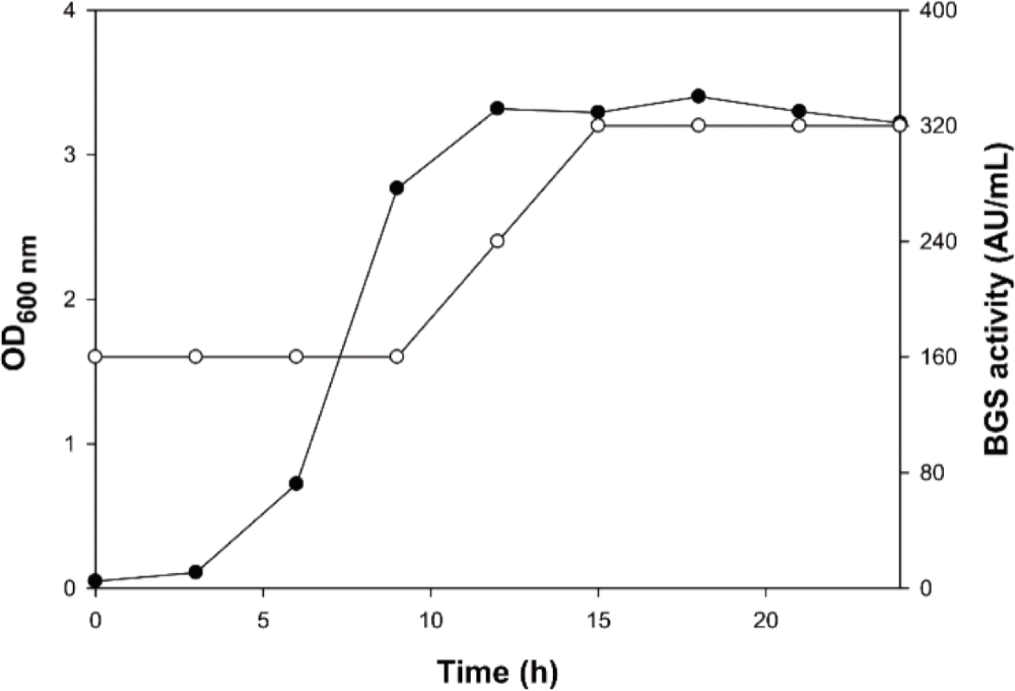
L. mesenteroides K748의 생육 곡선에 따른 BGS 활성은 Fig. 1에 나타내었다. 배양 3시간부터 대수 증식이 시작되었고, 배양 9시간 이후에 정지기에 도달하였다. BGS 활성은 한천배지확산법(Kouya et al., 2007)을 이용해 확인한 결과, 배양 0시간부터 대수 증식기까지 160 AU/mL 활성을 유지하였다. 배양 0시간부터 BGS 활성을 나타낸 것은 배양상등액에 포함된 MRS 배지 성분에 의한 B. longum의 생육 증가 효과일 가능성과 전배양에서 과량 생산된 BGS가 이미 포함되어 있었을 가능성이 있으나, 정확한 확인을 위해서는 추가적인 연구가 필요하다. 정지기인 배양 12시간 후부터 BGS 활성이 증가하여 배양 15시간 이후부터 24시간까지 320 AU/mL로 동일한 BGS 활성을 유지하였다. 배양 0시간에서 나타난 활성이 MRS 성분에 의한 B. longum의 생육 증가 효과라고 하더라도 배양 15시간 이후에 BGS 활성이 두 배 증가한 것은 L. mesenteroidesB. longum의 생육을 증가시키는 BGS를 생산했다는 것을 보여준다. Propionibacterium shermanii PZ-3의 경우, 대수기에서 BGS 활성이 증가하였고, 정지기 이후 BGS 활성이 유지되었으며(Kouya et al., 2008), Bacillus subtilis C-3102의 경우는 총균수 및 포자수가 증가하기 시작할 때, BGS 활성이 증가했으며, 정지기 이후에 배양 시간이 길어져도 BGS 활성은 유지되었다고 보고되었다(Hatanaka et al., 2020). 이와 유사하게, L. mesenteroides K748의 성장에 따른 BGS 활성도 대수기 후반 및 정지기 초반에 증가하기 시작하며, 정지기 이후부터 일정하게 유지되었고, 배양 시간이 24시간 이상으로 늘어나도 BGS 활성의 지속적 증가는 없을 것으로 예상된다.

rsr-5-2-72-g1
Fig. 1. Growth and BGS activity of L. mesenteroides K748 in MRS broth at 30°C. ●, growth curve measured at 600 nm; ○, BGS activity was measured using culture supernatant and the agar diffusion method.
Download Original Figure

또한, L. mesenteroides K748을 위와 같은 조건에서 혐기적으로 배양하며 3시간마다 상등액을 회수하여 BGS 활성을 확인한 결과, 최대 320 AU/mL의 활성으로 호기적 조건에서의 최대 BGS 활성과 동일하게 나타났다(데이터 불표시). Weisella paramesenteroides CJNU 0480의 경우 호기적 배양보다 혐기적 배양에서 BGS 생산량이 더 높았고 (Eom, 2012), Propionibacterium acidipropionici JCM 6432 및 P. jensenii JCM 6433의 경우는 호기적 배양에서 더 높은 BGS 생산량을 나타냈다(Kouya et al., 2007). 이와 같이, 균주마다 최대 BGS 활성을 나타내는 생육 조건이 다르기 때문에, L. mesenteroides K748의 최대 BGS 생산을 위한 최적 조건에 대한 추가 연구가 필요할 것으로 보인다.

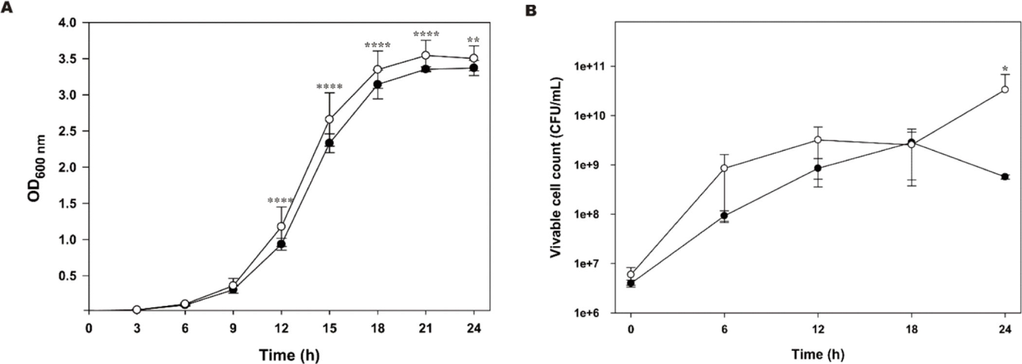
L. mesenteroides K748 배양 상등액 첨가가 B. longum KCTC 3128의 성장에 영향을 미치는지 확인하기 위해 배양액의 흡광도와 생균수를 측정하였으며, 그 결과는 Fig. 2와 같다. L. mesenteroides K748 배양 상등액을 첨가하였을 때, 흡광도 측정에서 9시간까지는 유의적인 차이를 나타내지 않았으나, 12시간 이후로는 대조군과 비교하였을 때 유의적으로 생육이 증가한 것으로 나타났다. L. mesenteroides K748 배양 상등액을 첨가 효과를 생균수 측정으로 확인한 결과는 하였을 때는 6시간, 12시간, 24시간에서 대조군에 비해 높은 생균수를 나타내었지만, 24시간에서만 유의적인 차이를 나타내었다. 24시간에서 대조군의 생균수는 줄어든 것으로 미루어 보아 B. longum이 사멸기에 접어든 것을 확인할 수 있는 반면, L. mesenteroides K748 배양 상등액을 첨가하였을 때는 24시간에서 생균수가 오히려 늘어난 것을 확인할 수 있다. 이는 L. mesenteroides K748이 생산하는 BGS 활성으로 B. longum의 대수기가 연장된 것으로 추정할 수 있다. 생균수 측정했을 때 6시간, 12시간에서 유의적인 차이는 보이지 않았지만 L. mesenteroides K748 배양 상등액을 첨가했을 때 B. longum의 생균수가 증가한 것은, 유청에서 배양한 L. casei LP1 균주의 배양 상등액을 이용하여 Bifidobacteirum 균주의 성장을 보았을 때 대조군과의 성장 차이가 배양 12시간 이전부터 나타났던 것과 유사한 결과라 할 수 있다(Kang et al., 2015).

rsr-5-2-72-g2
Fig. 2. BGS activity of cell-free supernatants of L. mesenteroides K748 for B. longum. (A) Growth curve from optical densities measurements. (B) Growth curve from viable cell count methods. B. longum was inoculated anaerobically in 37°C RCM broth. ●, B. longum KCTC 3128 only; ○, B. longum KCTC 3128 plus K748 cell free supernatant. Two-way ANOVA followed by uncorrected Fisher's least significant difference (LSD) multiple comparisons test, *, p<0.05, **, p<0.01, ****, P<0.0001.
Download Original Figure
BSG의 HPLC 분석

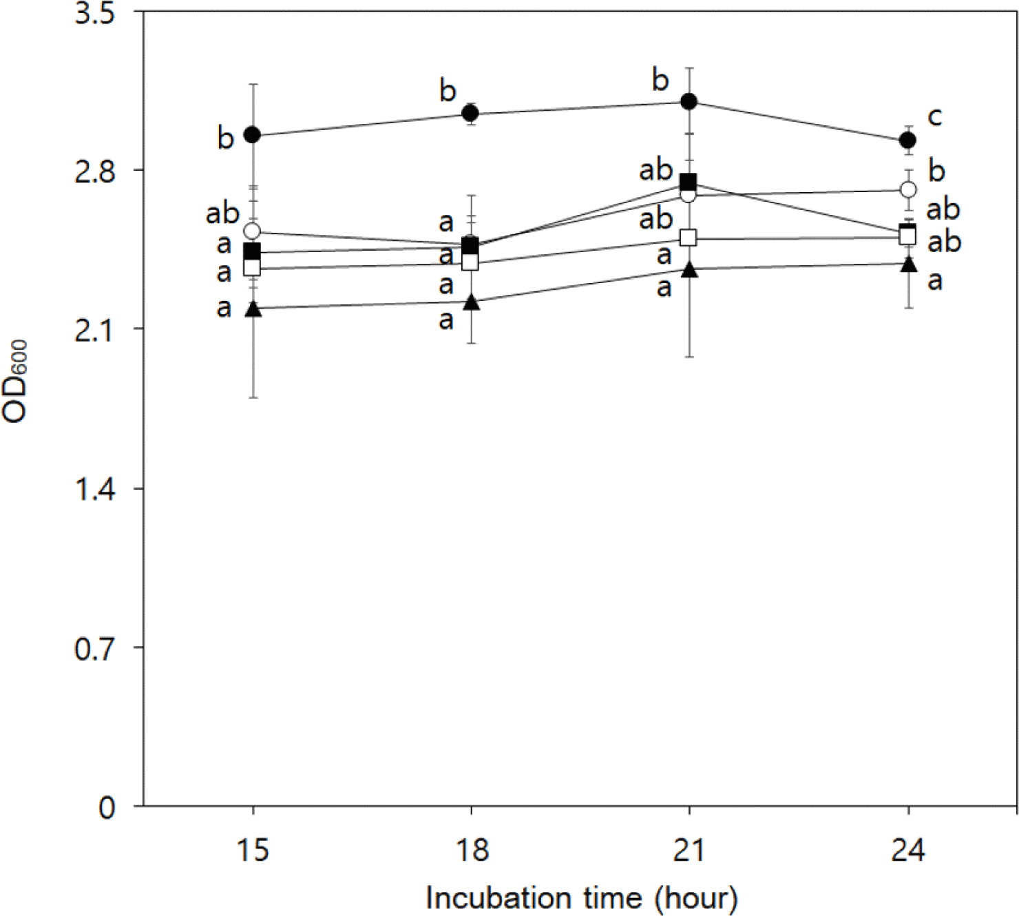
L. mesenteroides K748의 배양 상등액을 hexane, ethyl acetate, butanol의 순서대로분획 및 농축하여, 3가지 용매별 추출물을 각각 첨가하여 B. longum KCTC 3128의 성장 증가 효과를 확인하였다 (Fig. 3). Hexane과 butanol 추출물 첨가군에서는 무첨가군과 비교하였을 때 15시간과 18시간에서는 유의적인 차이를 나타내지 않았으나, 21시간과 24시간에는 유의적으로 증가하였다. 그러나 농축하지 않은 배양 상등액을 첨가한 경우와 비교하였을 때 15시간과 24시간에 유의적으로 흡광도가 낮게 나타나 BGS의 농축효과가 없는 것으로 사료된다. 반면에 ethyl acetate 추출물을 첨가하였을 때는 모든 시간대에서 농축하지 않은 배양 상등액을 첨가한 경우와 비교하였을 때 유의적으로 가장 높은 흡광도를 나타내어 BGS의 농축효과가 있는 것으로 확인되었다.

rsr-5-2-72-g3
Fig. 3. BGS activity of organic solvent extracts for B. longum. Each organic solvent extract was prepared from the cell-free supernatant of L. mesenteroides K748. B. longum was inoculated anaerobically in 37°C RCM broth. ▲, B. longum KCTC 3128 only; ○, B. longum KCTC 3128 plus K748 cell-free supernatant; ■, B. longum KCTC 3128 plus hexane extract of K748 cell-free supernatant; ●, B. longum KCTC 3128 plus ethyl acetate extract of K748 cell-free supernatant; □, B. longum KCTC 3128 plus butanol extract of K748 cell-free supernatant. IBM SPSS statistics program was used to analyze the results. Means with different letters are significantly different at p<0.05 by Duncan’s multiple range test.
Download Original Figure

한천배지확산법 (Kouya et al., 2007)을 통한 용매별 추출물의 BGS 활성 측정 결과는 Table 5와 같다. L. mesenteroides K748 상등액 100 mL를 분획 및 감압 농축 후, ddH2O를 이용해 5 mL로 정용하여 실험에 사용하였으며, hexane, ethyl acetate, butanol 추출물의 BGS 활성은 각각 160 AU/mL, 2,560 AU/mL, 640 AU/mL로 나타났다. 이를 통해 ethyl acetate 추출물의 BGS 활성이 가장 높은 것으로 확인되었고, ethyl acetate 추출물을 HPLC 분석에 사용하였다.

Table 5. Comparison of BGS activity of cell-free supernatant extracts by solvent
Sample Volume (mL) Specific activity (AU/mL)1) Purification fold Total activity (AU)2) Recovery (%)
K748 supernatant 100 320 - 32,000 -
Hexane ext. 5 160 0.25 800 2.5
Ethyl acetate ext. 5 2,560 8 12,800 40
Butanol ext. 5 640 2 3,200 10

1) The activity per 1 mL sample is defined as 20 AU.

2) Volume (mL) * Specific activity (AU/mL).

Download Excel Table

Propionibacterium freudenreichii 7025 균주가 생산하는 ACNQ의 경우, ethyl acetate에서 추출되었고(Mori et al., 1997), P. freudenreichii ET-3 균주가 생산하는 DHNA의 경우도 ethyl acetate에서 추출하였다는 결과가 보고되었다(Isawa et al., 2002). DHNA는 김치, 막걸리, 치즈 등과 같은 발효 식품 유래 유산균에서 분리한 연구가 다수 보고된 바 있기 때문에, 이를 토대로 L. mesenteroides K748이 생산하는 BGS 물질이 DHNA일 가능성이 높다고 추측되어 DHNA를 표준물질로 사용하였다.

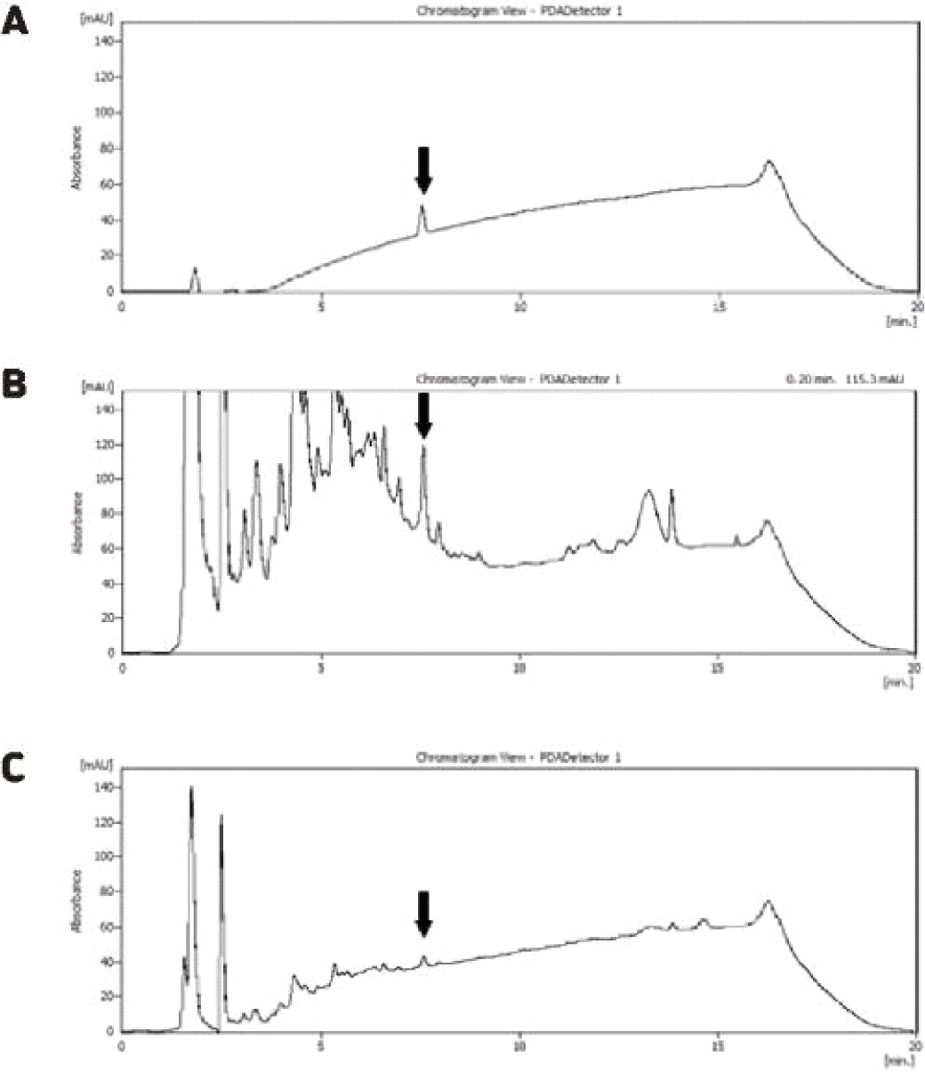
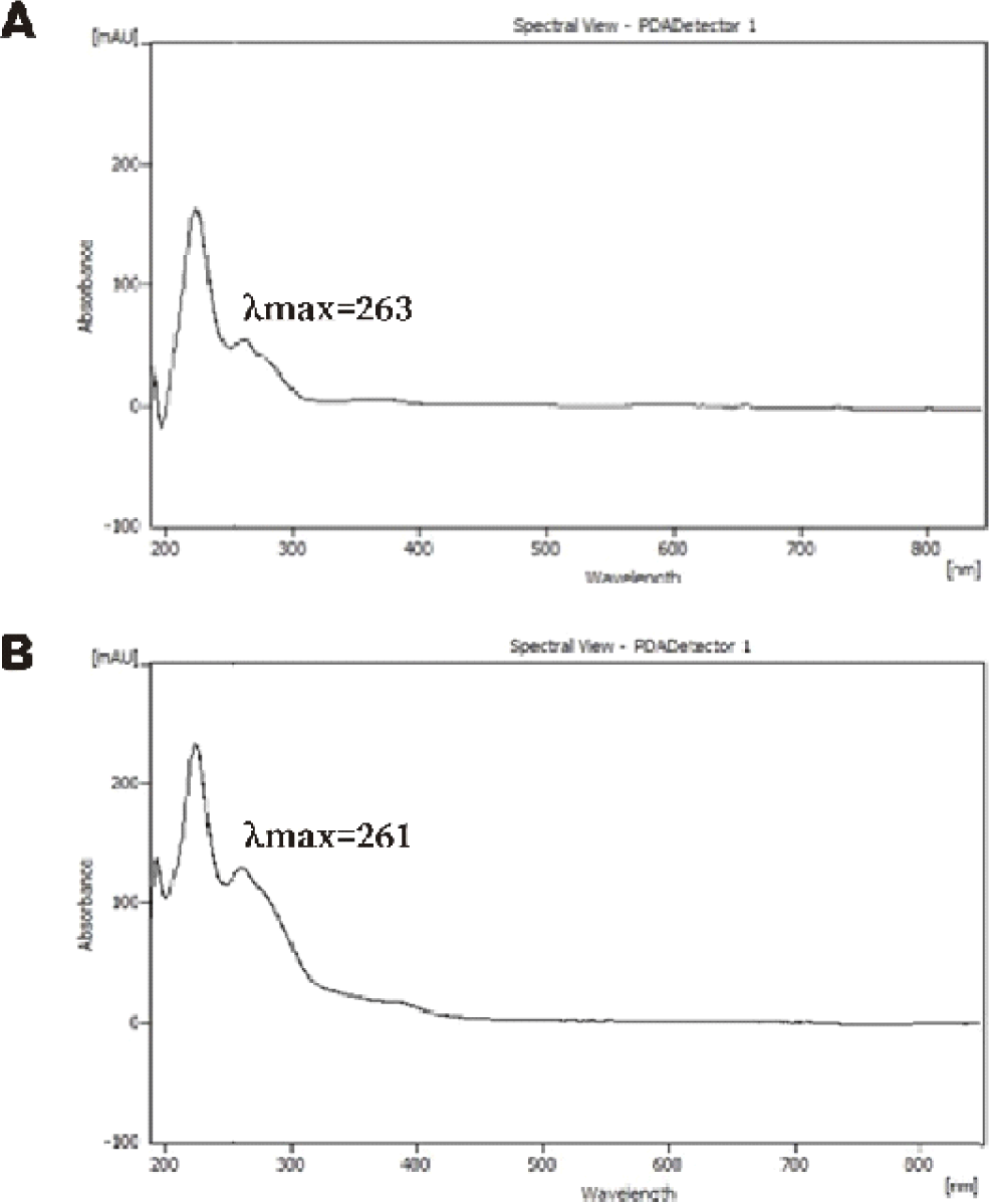
L. mesenteroides K748 상등액의 ethyl acetate 추출물을 HPLC를 이용해 분석한 결과는 Fig. 4와 같다. 표준물질인 DHNA와 비교하여 ethyl acetate 추출물과 10배 희석한 ethyl acetate 추출물에서 모두 동일하게 7.5분의 retention time을 가지는 피크를 확인하였다. 표준물질을 이용하여 작성한 검량곡선으로부터 계산한 ethyl acetate 추출물의 DHNA의 농도는 0.43 μmole이었다. 해당 피크의 UV spectrum 결과는 Fig. 5와 같으며, 254 nm에서 측정하였고, DHNA와 ethyl acetate 추출물에서 유사한 스펙트럼을 확인하였다. DHNA와 ethyl acetate 추출물의 최대흡수파장(λmax)은 263 nm 및 261 nm로 확인되었으며, 최대흡수파장에 차이가 있는 것은 정제되지 않은 ethyl acetate 추출물에 포함된 다른 물질이 영향이 미친 것으로 추측된다. Ethyl acetate 추출물의 BGS 활성 물질이 DHNA임을 명확히 하기 위해 LC-MS 분석을 진행하였다.

rsr-5-2-72-g4
Fig. 4. HPLC chromatogram of cell-free supernatant extracted with ethyl acetate. A, standard DHNA (2 ppm); B, ethyl acetate extract of L. mesenteroides K748 cell-free supernatant; C, 1/10 dilution sample B.
Download Original Figure
rsr-5-2-72-g5
Fig. 5. HPLC-UV spectrum of cell-free supernatant extract with ethyl acetate. Measurement was carried out at UV254 nm. A, standard DHNA (2 ppm); B, ethyl acetate extract of L. mesenteroides K748 cell-free supernatant.
Download Original Figure
BSG의 LC-MS 분석

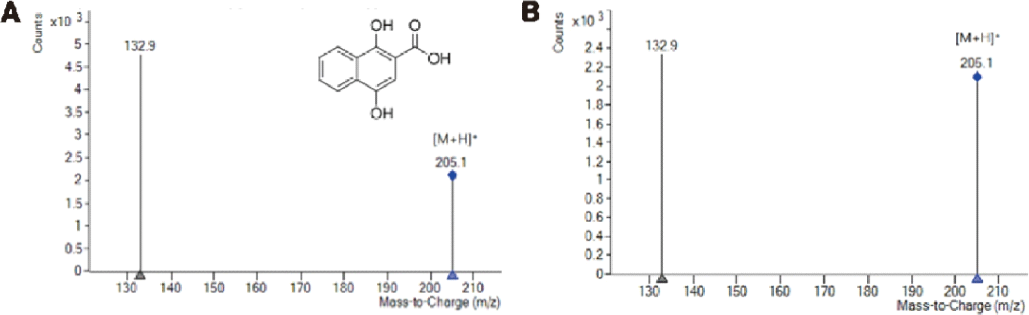
L. mesenteroides K748이 생산하는 BGS가 DHNA와 동일 물질인지 확인하기 위해 균주 상등액의 ethyl acetate 추출물을 LC-MS 분석하였다. DHNA 표준물질과 추출물 모두 4.4분대의 retention time에서 피크를 나타냈으며, 해당 피크의 MS 스펙트럼 결과는 Fig. 6과 같다. 양이온화 모드로 확인한 DHNA와 ethyl acetate 추출물에 포함된 물질의 [M+H]+ 이온은 m/z 205.1로 나타났으며, 이를 통해, ethyl acetate 추출물의 BGS 활성 물질이 204.1의 분자량을 가지는 DHNA와 동일함을 확인하였다. 최종적으로, L. mesenteroides K748이 생산하는 BGS 활성 물질은 DHNA로 추정된다.

rsr-5-2-72-g6
Fig. 6. LC-MS chromatogram of cell-free supernatant extract with ethyl acetate. Measurement was carried out at UV260 nm. A, standard DHNA (0.2 ppm); B, ethyl acetate extract of L. mesenteroides K748 cell-free supernatant.
Download Original Figure

DHNA는 메나퀴논(비타민 K2)의 중간 전구체이며, 메나퀴논은 박테리아 호흡계에서 전자전달제로 작용하며 동물성 제품 및 발효 식품에서 주로 발견되는 것으로 알려져 있다(Chen et al., 2013; Myneni and Mezey, 2017). DHNA가 첨가된 포뮬라를 노인 환자가 섭취했을 때, 분변에서 Bifidobacterium의 생균수가 유의적으로 증가하였고, 장내 미생물 균총을 개선할 수 있다는 보고가 있었다(Nagafuchi et al., 2015). DHNA는 BGS 역할 외에도 Helicobacter pylori의 성장 억제(Nagata et al., 2010), 대장염을 가진 마우스에서 Lactobaillus 및 Enterobacteriaceae의 회복 및 조직학적 손상 개선(Okada et al., 2006), 전염증성 사이토카인 조절을 통한 염증 완화(Okada et al., 2013) 등 생리활성 효과를 나타내는 것으로 알려져 있다.

하지만, 본 연구에서 L. mesenteroides K748의 DHNA 생산량은 미량으로 확인되었고, 추가로 DHNA의 생산성을 높이기 위한 연구가 필요할 것으로 보인다. DHNA의 생합성에는 E. coli에서 밝혀진 바에 의하면 menF, menD, menH, menC, menE, menB, menI 유전자가 관여한다고 알려져 있다(Chen et al., 2013). NCBI (http://www.ncbi.nlm.nih.gov/)의 BLAST (blastp) 프로그램을 이용하여 표준균주인 L. mesenteroides subsp. mesenteroides ATCC 8293의 DHNA 생합성 유전자 존재 여부를 확인한 결과, menI를 제외한 생합성 유전자를 보유하고 있음을 알 수 있었다. BGS 활성은 확인되었으 나 DHNA가 검출되지 않은 L. mesenteroides 균주에 DHNA-CoA synthase에 해당하는 menB 유전자를 과발현시켰을 때 DHNA의 생산이 유도된다는 보고가 있다(Eom and Moon, 2015). 따라서 L. mesenteroides K748이 가지는 DHNA 생산에 관여하는 유전자를 분석하고, 이를 활용한다면 DHNA 생산량을 증가시킬 수 있을 것으로 사료된다.

Ⅳ. 요 약

Bifidobacterium 속은 장내 유익균으로 알려져 있으며, Bifidobacterium의 일부 균주는 프로바이오틱스로 사용되고 있다. Bifidobacterium의 성장을 촉진하는 Bifidobacterium 성장 자극제(BGS)를 생산하는 유산균을 찾기 위해 김치에서 분리한 733개 균주를 대상으로 BGS 스크리닝을 실시하여 320 AU/mL로 가장 높은 BGS 활성을 보인 K748 균주를 선발하였다. 이 균주는 16S rDNA 염기서열 분석으로 L. mesenteroides로 확인되었고, 균주의 이름은 L. mesenteroides K748로 명명하였다. BGS 활성은 MRS 배지에서 배양 후 12시간부터 증가하여 15시간에 최대 활성(320 AU/mL)에 도달하였고, 이후에도 그 활성을 유지했다. L. mesenteroides K748 배양 상등액의 ethyl acetate 추출물은 B. longum의 성장을 촉진했으며, 이 추출물을 사용한 HPLC는 1,4-dihydroxy-2-naphthoic acid (DHNA)와 동일한 체류 시간(7.5분)을 가진 피크를 나타냈다. LC-MS 분석 결과, L. mesenteroides K748의 배양 상등액의 에틸아세테이트 추출물은 분자량이 204.1로 DHNA와 동일하게 나타났다. 결론적으로 L. mesenteroides K748은 Bifidobacterium의 성장을 촉진하는 DHNA를 생산한다는 것을 확인하였다.

Ⅴ. 참고문헌

1.

Aguilar-Toalá JE, Garcia-Varela R, Garcia HS, Mata-Haro V, González-Córdova AF, Vallejo-Cordoba B, et al. Postbiotics: An evolving term within the functional foods field. Trends Food Sci 75:105-114.

2.

Chen M, Ma X, Chen X, Jiang M, Song H, Guo Z. Identification of a hotdog fold thioesterase involved in the biosynthesis of menaquinone in Escherichia coli. J Bacteriol 195:2768-2775.

3.

Chin HS, Breidt F, Fleming HP, Shin W, Yoon S. Identification of predominant bacterial isolates from the fermenting kimchi using its-PCR and partial 16s RDNA sequence analyses. J Microbiol Biotechnol 16:68-76.

4.

Choi N-Y, Shin H-S. Effect of oligosaccharides and inulin on the growth and viability of Bifidobacteria in skim milk. Food Sci Biotechnol 15:543-548.

5.

Chun BH, Kim KH, Jeon HH, Lee SH, Jeon CO. Pan-genomic and transcriptomic analyses of Leuconostoc mesenteroides provide insights into its genomic and metabolic features and roles in kimchi fermentation. Sci Rep 7:11504.

6.

Doré J, Blottière H. The influence of diet on the gut microbiota and its consequences for health. Curr Opin Biotechnol 32:195-199.

7.

Eom JE. Isolation and application of lactic acid bacteria producing 1,4-dihydroxy-2- naphthoic acid. Master thesis. Chungju University.

8.

Eom JE, Moon GS. Construction of a recombinant Leuconostoc mesenteroides CJNU 0147 producing 1,4-dihydroxy-2-naphthoic acid, a bifidogenic growth factor. Kor J Food Sci Anim Resour 35:867-873.

9.

Eom JE, Kwon SC, Moon GS. Detection of 1,4-dihydroxy-2-naphthoic acid from commercial makgeolli products. Prev Nutr Food Sci 17:83-86.

10.

Gourbeyre P, Denery S, Bodinier M. Probiotics, prebiotics, and synbiotics: Impact on the gut immune system and allergic reactions. Leukoc Biol 89:685-695.

11.

Hatanaka M, Morita H, Aoyagi Y, Sasaki K, Sasaki D, Kondo A, et al. Effective bifidogenic growth factors cyclo-Val-Leu and cyclo-Val-ile produced by Bacillus subtilis C-3102 in the human colonic microbiota model. Sci Rep 10:7591.

12.

Iraporda C, Rubel IA, Manrique GD, Abraham AG. Influence of inulin rich carbohydrates from jerusalem artichoke (Helianthus tuberosusL.) tubers on probiotic properties of Lactobacillus strains. LWT 101:738-746.

13.

Isawa K, Hojo K, Yoda N, Kamiyama T, Makino S, Saito M, et al. Isolation and identification of a new bifidogenic growth stimulator produced by Propionibacterium freudenreichii ET-3. Biosci Biotechnol Biochem 66:679-681.

14.

Jung HS, Kim KR, Kim KH, Kim H-M, Lim S, Chung MJ, et al. Investigation on human milk oligosaccharide utilization and vitamin B biosynthesis by Bifidobacterium strains isolated from infant feces. Kor J Microbiol 56:297-306.

15.

Jung JY, Lee SH, Lee HJ, Seo HY, Park WS, Jeon CO. Effects of Leuconostoc mesenteroides starter cultures on microbial communities and metabolites during kimchi fermentation. Int J Food Microbiol 153:378-387.

16.

Kang JE, Kim TJ, Moon GS. A novel Lactobacillus casei LP1 producing 1,4- dihyroxy-2-naphthoic acid, a bifidogenic growth stimulator. Prev Nutr Food Sci. 20:78-81.

17.

Khanna S, Tosh PK. A clinician's primer on the role of the microbiome in human health and disease. Mayo Clin Proc 89:107-114.

18.

Kim JM. Roles of enteric microbial composition and metabolism in health and diseases. Kor J Gastroenterol 62:191-205.

19.

Kim MJ, Ku S, Kim SY, Lee HH, Jin H, Kang S, et al. Safety evaluations of Bifidobacterium bifidum BGN4 and Bifidobacterium longum Bori. Int J Mol Sci 19.

20.

Kim TJ, Moon GS. Antimicrobial activity of Lactobacillus plantarum LP2 against Helicobacter pylori. J Food Hyg Saf 30:372-375.

21.

Kim Y-H, Kim H-Z, Kim J-Y, Choi T-B. Strain improvement of Leuconostoc mesenteroides as a acid-resistant mutant and effect on kimchi fermentation as a starter. Microbiol Biotechnol Lett 33:41-50.

22.

Knysh OV. Bifidogenic properties of cell-free extracts derived from probiotic strains of Bifidobacterium bifidum and Lactobacillus reuteri. Regul Mech Biosyst 10:124-128.

23.

Kouya T, Misawa K, Horiuchi M, Nakayama E, Deguchi H, Tanaka T, et al. Production of extracellular bifidogenic growth stimulator by anaerobic and aerobic cultivations of several propionibacterial strains. J Biosci Bioeng 103:464-471.

24.

Kouya T, Tobita K, Horiuchi M, Nakayama E, Deguchi H, Tanaka T, et al. Production of extracellular bifidogenic growth stimulator (BGS) from Propionibacterium shermanii using a bioreactor system with a microfiltration module and an on-line controller for lactic acid concentration. J Biosci Bioeng 105:184-191.

25.

Lee KW, Kim GS, Baek AH, Hwang HS, Kwon DY, Kim SG, et al. Isolation and characterization of kimchi starters Leuconostoc mesenteroides PBIO03 and Leuconostoc mesenteroides PBIO104 for manufacture of commercial kimchi. J Microbiol Biotechnol 30:1060-1066.

26.

Lee SH, Chang HC. Isolation of antifungal activity of Leuconostoc mesenteroides ta from kimchi and characterization of its antifungal compounds. Food Sci Biotechnol 25:213-219.

27.

Meyer D, Stasse-Wolthuis M. The bifidogenic effect of inulin and oligofructose and its consequences for gut health. Eur J Clin Nutr 63:1277-1289.

28.

Mori H, Sato Y, Taketomo N, Kamiyama T, Yoshiyama Y, Meguro S, et al. Isolation and structural identification of bifidogenic growth stimulator produced by Propionibacterium freudenreichii. J Dairy Sci 80:1959-1964.

29.

Myneni VD, Mezey E. Regulation of bone remodeling by vitamin K2. Oral Dis 23:1021-1028.

30.

Nagafuchi S, Yamaji T, Kawashima A, Saito Y, Takahashi T, Yamamoto T, et al. Effects of a formula containing two types of prebiotics, bifidogenic growth stimulator and galacto-oligosaccharide, and fermented milk products on intestinal microbiota and antibody response to influenza vaccine in elderly patients: A randomized controlled trial. Pharmaceuticals (Basel) 8:351-365.

31.

Nagata K, Inatsu S, Tanaka M, Sato H, Kouya T, Taniguchi M, et al. The bifidogenic growth stimulator inhibits the growth and respiration of Helicobacter pylori. Helicobacter 15:422-429.

32.

Nagpal R, Yadav H, Marotta F. Gut microbiota: The next-gen frontier in preventive and therapeutic medicine? Front Med (Lausanne) 1:15.

33.

Okada Y, Tsuzuki Y, Miyazaki J, Matsuzaki K, Hokari R, Komoto S, et al. Propionibacterium freudenreichii component 1,4-dihydroxy-2-naphthoic acid (DHNA) attenuates dextran sodium sulphate induced colitis by modulation of bacterial flora and lymphocyte homing. Gut 55:681-688.

34.

Okada Y, Tsuzuki Y, Narimatsu K, Sato H, Ueda T, Hozumi H, et al. 1,4-Dihydroxy-2-naphthoic acid from Propionibacterium freudenreichii reduces inflammation in interleukin-10-deficient mice with colitis by suppressing macrophage- derived proinflammatory cytokines. J Leukoc Biol 94:473-480.

35.

Pandey KR, Naik SR, Vakil BV. Probiotics, prebiotics and synbiotics- a review. J Food Sci Technol 52:7577-7587.

36.

Paray BA, Rather IA, Al-Sadoon MK, Fanar Hamad AS. Pharmaceutical significance of Leuconostoc mesenteroides KS-TN11 isolated from nile tilapia, Oreochromis niloticus. Saudi Pharm J 26:509-514.

37.

Park MS, Ji GE. Research trends in Bifidobacterium. KSBB J 25:319-329.

38.

Piqué N, Berlanga M, Miñana-Galbis D. Health benefits of heat-killed (tyndallized) probiotics: An overview. Int J Mol Sci 20.

39.

Sakai Y, Seki N, Hamano K, Ochi H, Abe F, Masuda K, et al. Prebiotic effect of two grams of lactulose in healthy Japanese women: A randomised, double-blind, placebo-controlled crossover trial. Benef Microbes 10:629-639.

40.

Schleifer K-H. Phylum xiii. Firmicutes gibbons and murray 1978, 5. De Vos P, Garrity GM, Jones D, Krieg NR, Ludwig W, Rainey FA, et al., editors. Bergey’s manual® of systematic bacteriology: Volume three the firmicutes. Springer New York New York, NY. 2009.

41.

Sims IM, Tannock GW. Galacto- and fructo-oligosaccharides utilized for growth by cocultures of bifidobacterial species characteristic of the infant gut. Appl Environ Microbiol 86.

42.

Vitellio P, Celano G, Bonfrate L, Gobbetti M, Portincasa P, De Angelis M. Effects of Bifidobacterium longum and Lactobacillus rhamnosus on gut microbiota in patients with lactose intolerance and persisting functional gastrointestinal symptoms: A randomised, double-blind, cross-over study. Nutrients 11.

43.

Wong CB, Odamaki T, Xiao J-z. Beneficial effects of Bifidobacterium longum subsp. longum BB536 on human health: Modulation of gut microbiome as the principal action. J Funct Foods 54:506-519.